Nobel 2026: How to Give Nerve Cells a Light Switch
How an alga’s response to light became a tool for controlling nerve cells, and what optogenetics could mean for vision, hearing and drug discovery.
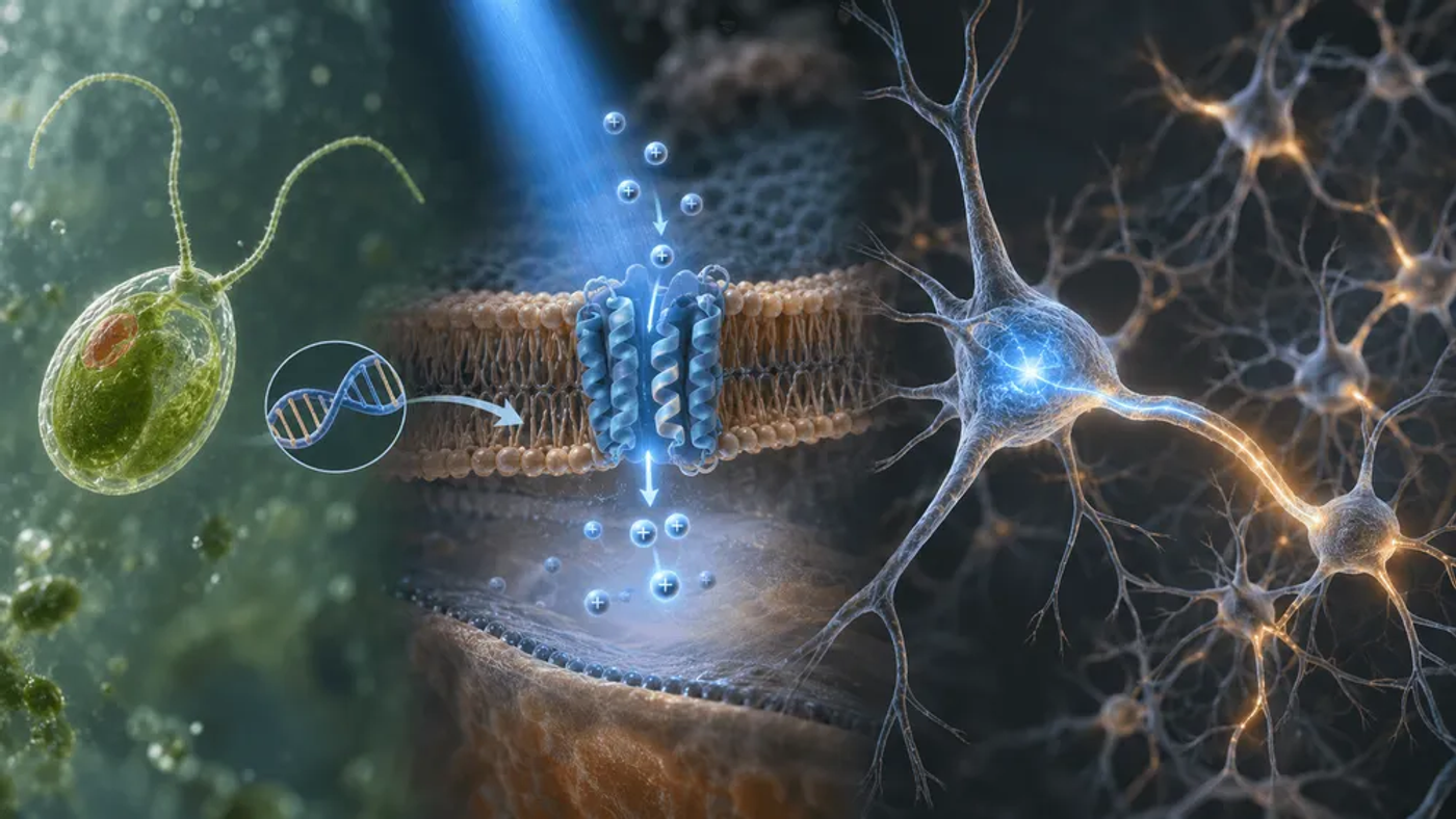
A green alga’s response to light helped scientists develop a switch for nerve cells. That work led to optogenetics, which earned Peter Hegemann, Georg Nagel and Karl Deisseroth the 2026 Nobel Prize in Physiology or Medicine.

By giving selected cells genes for light-sensitive proteins, researchers can activate or silence them with light. This lets them test what specific neurons do and explore ways to restore lost functions such as vision.
It started with an alga looking for light
Chlamydomonas is a single-celled green alga that uses light for photosynthesis. It can swim towards or away from light, which raised a small but important question. How does it turn incoming light into a signal that changes its behaviour?
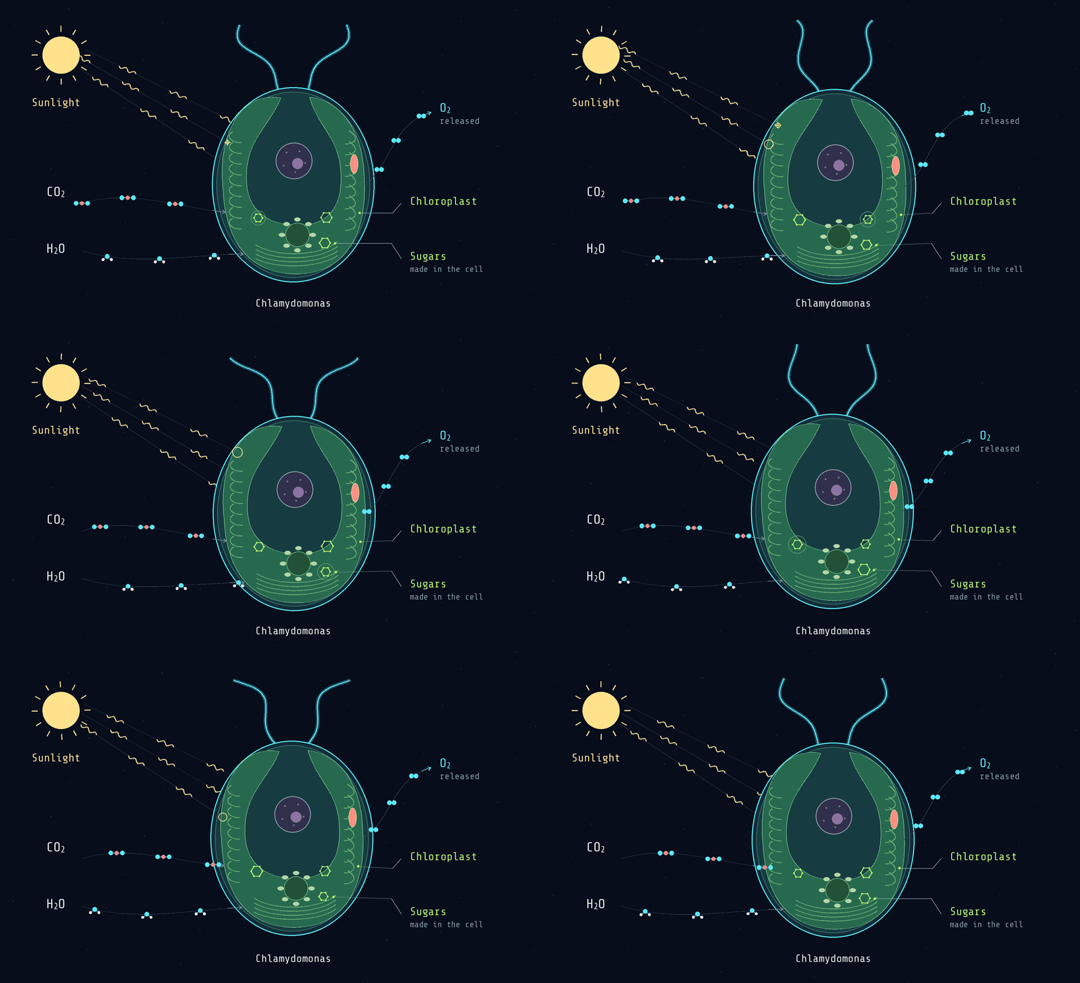
Hegemann and his collaborators investigated the proteins involved. In work with Nagel, Ernst Bamberg and other researchers, they identified and characterised channelrhodopsins: proteins that form light-controlled channels in a cell membrane.
The membrane separates the inside of a cell from its surroundings. A channel provides a route for charged particles, called ions, to cross it. In this case, light opens the route.
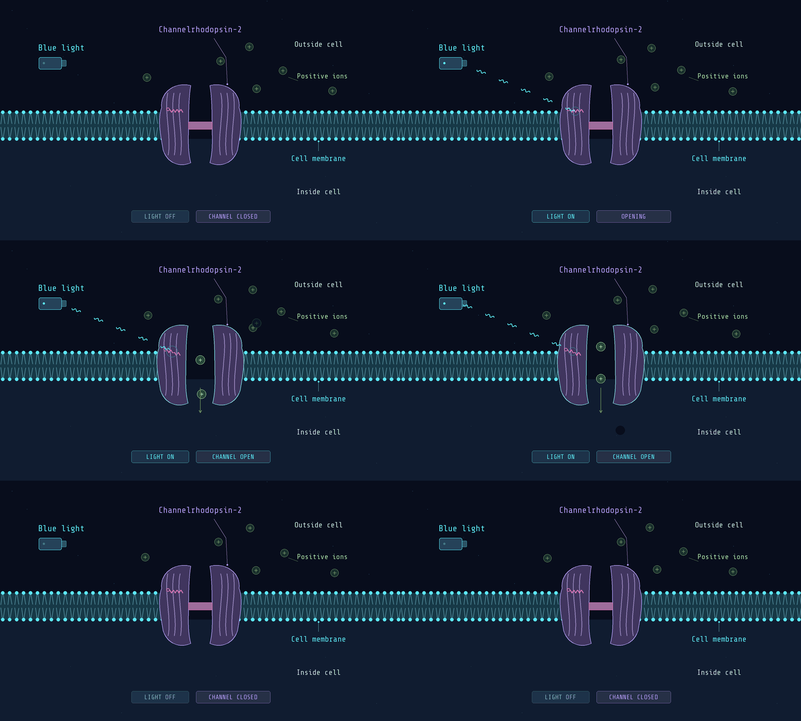
Researchers described channelrhodopsin-1 in 2002, followed by channelrhodopsin-2 in 2003. The latter lets positively charged ions cross a cell membrane when activated by light.
In 2005, Edward Boyden, Feng Zhang, Ernst Bamberg, Georg Nagel and Karl Deisseroth brought this mechanism into cultured mammalian neurons. Brief light pulses could trigger electrical signals with millisecond precision, turning an algal protein into a tool for controlling selected nerve cells. The Nobel recognises three scientists, but this breakthrough involved a wider group.
How do you give a cell a light switch?
Optogenetics combines three things: a light-sensitive protein, a way to get its genetic instructions into the right cells, and hardware that delivers the light.
First, choose the cells
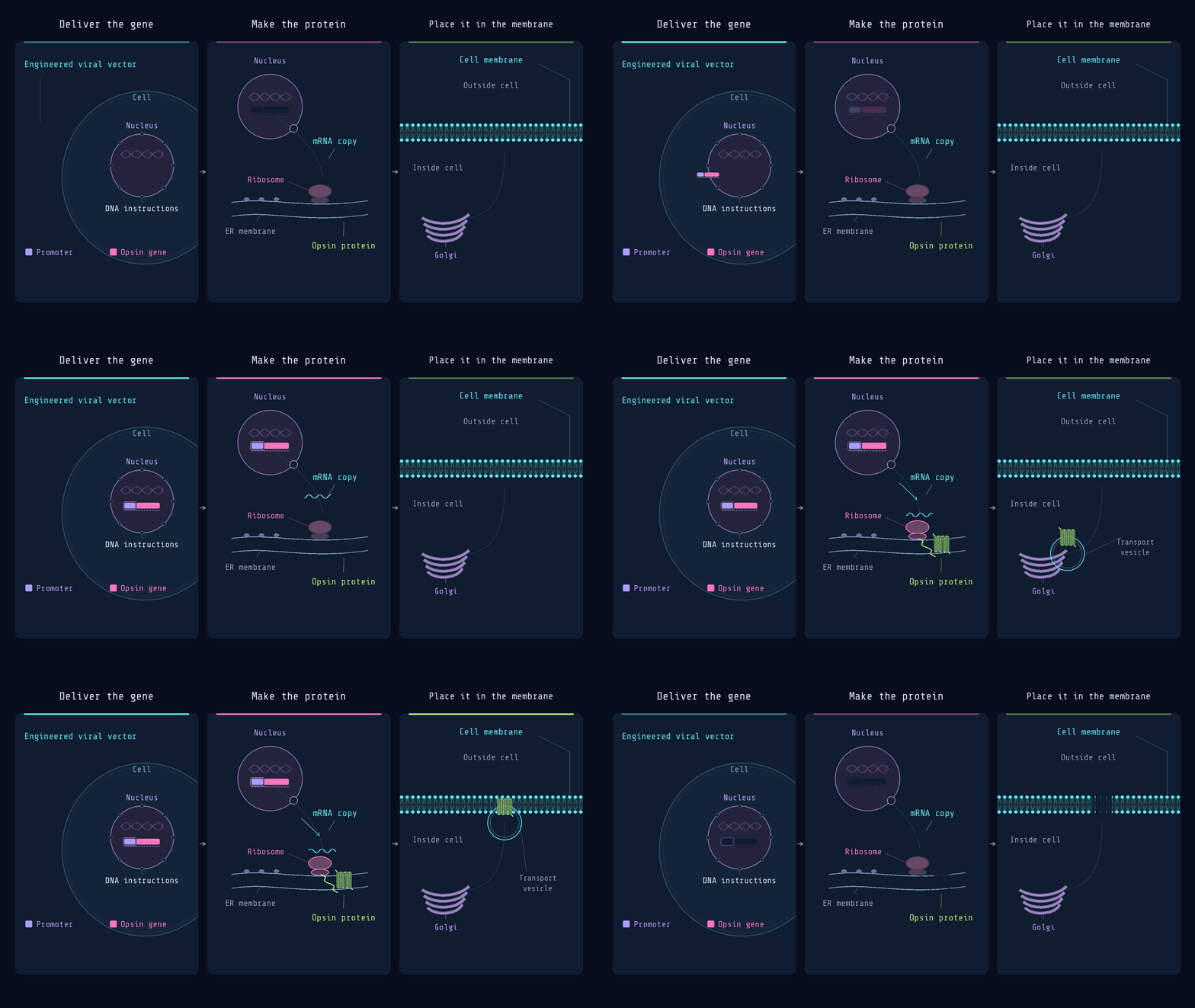
Researchers introduce a gene for an opsin, a light-sensitive protein, often using an engineered viral vector. Regulatory DNA helps control which cells produce it, although targeting is imperfect.
The cells then make the protein. For an ion-channel opsin, it sits in the cell membrane, where it can control ion movement.
Then, use light to change their electrical activity
Neurons send electrical signals through changes in the voltage across their membranes.
In channelrhodopsin-2, a molecule called retinal absorbs blue light. This changes the protein’s shape and opens its channel. Positively charged ions can enter, bringing the neuron closer to firing an electrical impulse.
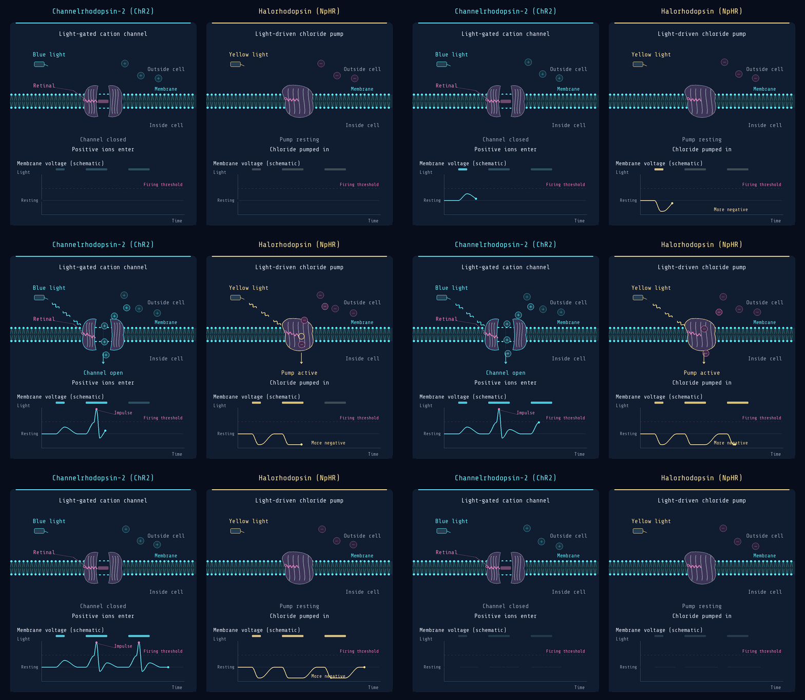
Other opsins suppress activity. Different proteins respond to different colours and at different speeds, giving researchers control over the timing and pattern of neural activity.
Finally, deliver the light and measure the result
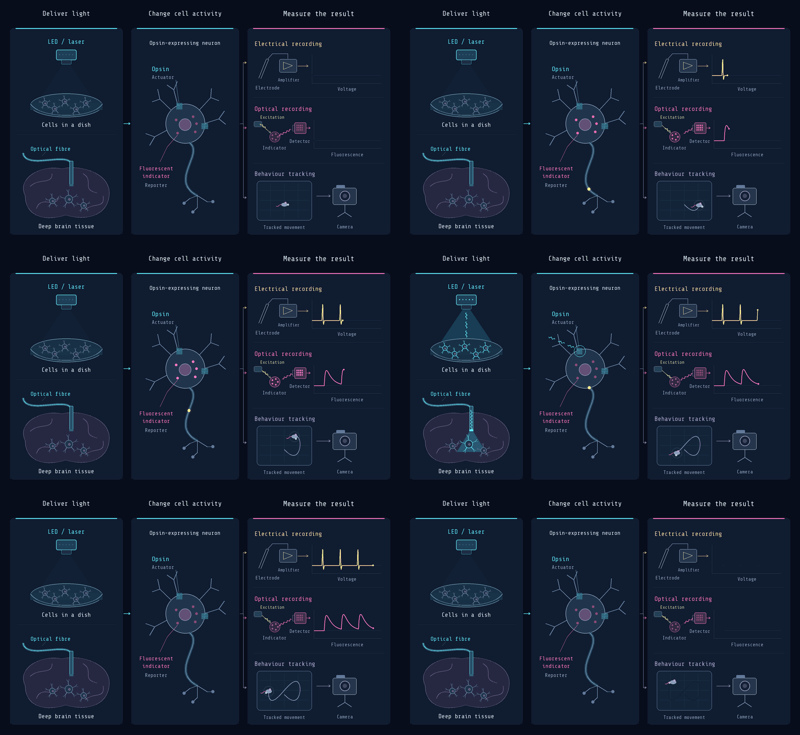
An LED or laser supplies the appropriate wavelength. Cells in a dish are relatively easy to illuminate; deep brain targets often require an implanted optical fibre.
Researchers then measure the response using electrodes, fluorescent sensors or changes in behaviour. The opsin controls activity, while a separate sensor or recording method measures what happens.
This engineered light sensitivity is what distinguishes optogenetics from ordinary light therapy.
Why this changed neuroscience, and could change treatment
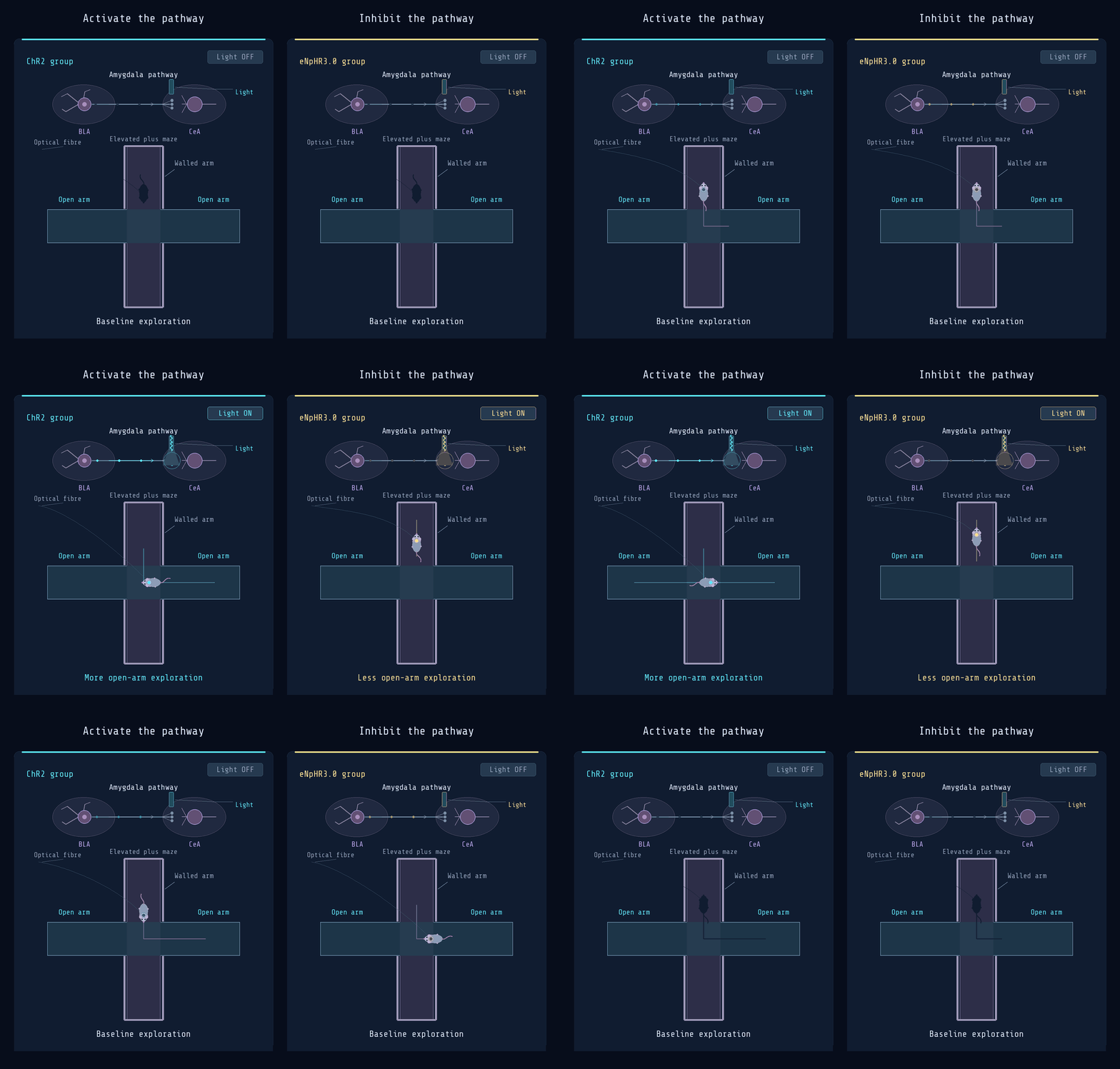
Optogenetics let researchers change fear- and anxiety-related behaviour by switching specific brain pathways on or off.
In a 2011 study, Kay Tye, Karl Deisseroth and colleagues activated a particular pathway in the amygdala, a brain region involved in fear and anxiety. Mice became more willing to explore exposed spaces they would normally avoid. Silencing the same pathway increased anxiety-like behaviour. These changes were rapid and reversible. The researchers measured behaviour, so “fear to calm” is a useful shorthand rather than a direct measurement of the animals’ mood.
A separate 2012 study showed control over fear-memory recall. Researchers labelled neurons active during fear learning, then reactivated them with light. The mice froze, a characteristic fear response, even in a different environment. This showed that activating a specific population of cells could trigger the behavioural expression of a learned memory.
Brain research and schizophrenia
Optogenetics lets researchers test whether specific neurons contribute to a behaviour. In a 2012 study, reactivating neurons labelled during fear learning could evoke a learned fear response in mice. Researchers also use it to investigate circuits implicated in schizophrenia, helping identify which changes in brain activity could become treatment targets.
Vision restoration
Optogenetic therapies aim to make surviving retinal cells detect light after the original photoreceptors have died. They use the cells that remain rather than regrowing lost ones.
In a 2021 report, one blind patient could locate and count some objects after treatment and training with light-stimulating goggles. This demonstrated partial visual function. Other approaches aim to work under ordinary environmental light.
Hearing restoration
Optical cochlear implants aim to stimulate light-sensitive auditory neurons more selectively than electrical implants. This could improve how different sound frequencies are distinguished, but both the gene therapy and implant must prove safe and useful in people.
Drug screening
Researchers stimulate laboratory-grown human neurons and measure how candidate drugs change their responses. Optogenetics becomes a tool for testing medicines; the eventual treatment does not need to involve light.
Technical constraints
Light weakens as it passes through tissue, and intense illumination can cause heating. Gene delivery must reach the intended cells and keep the opsin working safely over time. Implanted devices add further challenges.
For treatments, the decisive result is a sustained improvement in everyday function, not simply a measurable response to light.
Who is building on the discovery?
Selected companies applying optogenetics to experimental treatments, drug discovery and research tools. Public development status checked on 8 November 2026.
▣ Vision restoration
▣ Hearing restoration
▣ Drug discovery
▣ Research hardware
The field has developed in several connected directions. Protein engineering has produced opsins with different colours and speeds, including tools described in the 2014 work on Chronos and Chrimson. All-optical electrophysiology combines light-based stimulation with optical voltage measurements, allowing researchers to stimulate cells and read their responses.
Hearing is an interesting example of the engineering still required. Conventional cochlear implants stimulate auditory nerves electrically. Optical stimulation could potentially activate smaller groups of neurons more selectively, improving how different sound frequencies are represented. But it needs both a successful gene-delivery approach and a safe, durable implant. The potential advantage still has to translate into benefit for people.
What could happen next?
| Area | Current position | Next evidence needed | Potential outcome |
|---|---|---|---|
| Research & drug discovery | Light-controlled cells and optical recordings | More predictive human-cell models | Better tests for new medicines |
| Vision restoration | Clinical trials and regulatory review | Controlled benefit and long-term safety | Durable improvements in useful vision |
| Hearing restoration | Preclinical optical cochlear implants | Safe gene delivery and implant validation | More precise hearing stimulation |
Optogenetics may keep expanding as a research and drug-testing tool while its medical applications develop more unevenly. Vision restoration has a practical advantage because light can reach the retina, and clinical programmes are already testing whether surviving cells can provide useful sight. The next milestones are clearer evidence of benefit, long-term safety and treatment systems people can use comfortably.
Hearing restoration could follow if selective optical stimulation proves useful and the gene therapy and implant work reliably together. Deep-brain treatments face greater delivery challenges. The most convincing progress will come from a few well-defined applications where controlling the right cells produces a lasting improvement in what someone can do.